Every day, diligent researchers are making scientific breakthroughs that help change the way we understand and interact with the world. Much of the exploration that leads to these achievements occurs under a microscope — where the building blocks of life are magnified and their otherwise unseen properties made visible to the human eye.
Established in 2010, the annual Koch Institute Image Awards at the Massachusetts Institute of Technology pay homage to that process, celebrating “the extraordinary visuals produced through life sciences and biomedical research” at the world-renowned university.
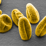
From a close-up look at mutated grains of pollen to a dazzling action shot of a cell dividing in two, scroll to view the 10 fascinating images that won this year’s awards, and read the interesting stories behind them.
Note: The image explanations have been provided via a Koch Institute Image Awards press release
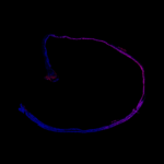
“A microneedle patch developed by the Hammond and Irvine Labs non-invasively samples biomarkers and immune cell populations in skin. By analyzing the cells and surrounding tissue, researchers aim to classify the immune signatures of various disease stages, including autoimmune skin conditions and hard to detect ovarian cancers. Highlighted here are immune cells (colored purple to represent gender equity and women’s health) collected by the needles’ seaweed-based coating (gray). The device is now being used in preclinical and clinical settings on three continents.”
Dancing With the Cells: Spotlight on Plasma Membrane
“The Manalis Lab studies the mechanical and biophysical changes that occur during cell division. In this experiment, researchers track the organization of the plasma membrane (red/yellow) as a live human adenocarcinoma cell divides in two. A lingering plasma bridge (center) contains remnants of DNA (blue) that failed to separate between the daughter cells. Such segregation errors can result in mutations that can initiate and drive cancer. Understanding how plasma membrane movement is regulated as normal and cancer cells divide could help explain how mutations arise and how to prevent them.”
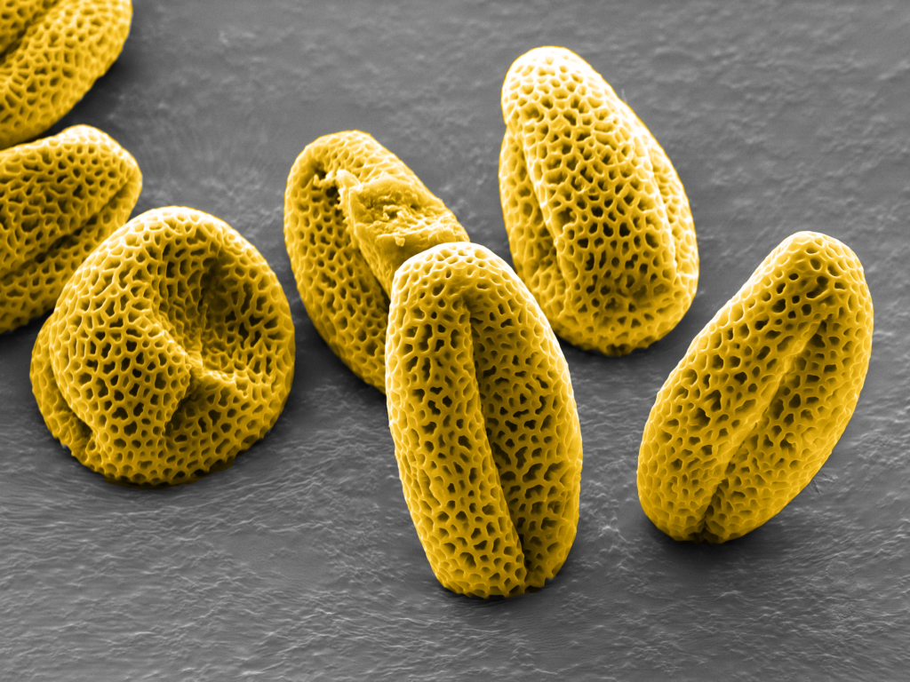
Grains of Truth: Evolution Shapes Cells
“The nuclear lamina is the innermost layer of a cell’s nuclear envelope — a protein meshwork that interacts with the genome and other proteins. Lack of lamina is lethal to plants and animals alike, yet the structure evolved independently in both. The Gehring Lab studies how the nuclear envelope regulates plant epigenetics and reproduction. The image shows pollen from the Arabidopsis thaliana mutant lacking three out of four lamina genes called CRWNs. These mutations cause inefficient reproduction and abnormal gene expression, as well as structural irregularities, most visible in the grains on the left.”
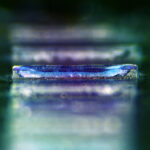
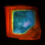
“This image of the distal colon shows how a highly focused radiation beam can be precisely targeted to induce DNA damage to nuclei in a region of interest (pink) without affecting the neighboring cells (blue). The Vander Heiden Lab is studying the effects of spatially targeted radiotherapy on metabolism as well as immune and stromal (support) cells in the tumor microenvironment. Understanding the molecular impacts of this approach will help physicians identify key pathways and therapeutic combinations to improve efficacy of radiotherapy in the clinic. For patients, the clear distinction between damaged and undamaged tissue offers hope for more strategic treatments with fewer side effects.”
Just Grow With It: Partnering with Patients to Create Desmoid Tumor Models
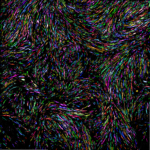
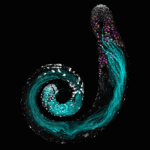
“Patients with rare tumors are partnering with Boehm Lab researchers to develop new cell models for drug testing and other applications. The team uses high throughput screening and computer assisted microscopy to find cell culture recipes that promote growth of donated cells. These pseudo-colored cells are taken from desmoid tumors, a rare form of sarcoma. Evidence of the model’s success is seen in the abundance of cells and the swirling growth pattern. The team is now expanding this patient-powered research to other understudied tumors, and sharing the models with researchers across the globe.”
Polymer Rising: A Shining Example for Global Vaccine Equity
“All that glitters… is not from cold storage! To promote global vaccine equity, Jaklenec and Langer Lab researchers are developing a micro-scale particle for long-term storage and one-shot delivery of mRNA vaccines in low-income communities where technical barriers limit access to cold-chain transportation. They took this image to visually assess how a polymer coating (pink) protects and stabilizes the dried mRNA vaccine (blue). Eventually, the container will be embedded in a dissolvable needle and injected into the body to release multiple doses of the active vaccine.”
Remodel Organism: Tissue Origami in a Fruit Fly Embryo
“As an organism transforms from a simple collection of cells into a complex system of life, the developing tissue undergoes extensive remodeling. The Martin Lab uses fruit fly embryos to study the impact of mechanical forces on cell behavior. On the left, nuclei in gray are linked by new cell junctures, marked in orange. The view on the right maps cell boundaries with randomly assigned colors to track them as they evolve. At center, a newly formed structure fold pulls the two sides inward. Collectively, these perspectives paint a dynamic picture of a cellular community working together to shape the organism.”
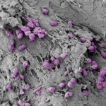
Shell Game: Microparticles Meet Immune Cells
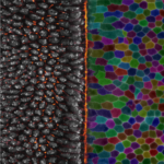
“Core shell microparticles developed in the Jaklenec/Langer Lab are designed to hold vaccines or immunotherapies and release them over days or months through controlled degradation of the encapsulating polymers. To determine whether immune cells in the skin will affect release timing, researchers first study the immune response to an unfilled microparticle after injection. This slice shows immune cells (green) on the interior surface of the polymer shell (red) beneath its biodegraded cap (blue/pink). Further classification of the cells will predict their interaction with the particles’ eventual cargo.”
Tail as Old as Time: Germ Cell Development in the Fruit Fly
“During development, Drosophila germ cells undergo incredible morphological changes to produce some of the longest sperm in the animal kingdom, ~2mm. In this fly testis, nuclei are marked in white. Germ cells begin their journey as stem cells (at the top). Continued division and maturation push cells away from this origin, eventually yielding mature sperm, shown in cyan. In between, magenta and yellow mark expression of RNAs essential for sperm development. The Yamashita Lab studies how every stage of germ cell development works together to ensure continued fertility from generation to generation.”
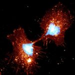
That Takes Nerve: Co-Culturing an Interest in Cancer Neuroscience
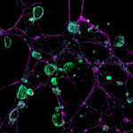
“Nerve cells are frequently overlooked in studies of the tumor microenvironment — which also includes blood vessels, immune cells, and many others — but they play a significant role in cancer patients’ survival and quality of life. Using genetically engineered mini-organs, the Jacks and Hwang Labs created a 3D model system where pancreatic cancer cells (green/cyan) interact with nerve cells (magenta). They seek to understand how the nervous system impacts tumor formation, growth, metastasis, and even drug resistance.”
Learn more about the Koch Institute Image Awards here.
RELATED: New Stamp-Sized Ultrasound Sticker Images Internal Organs, Could Stand in for Bulky Machines


Pingback: Revival do Movimento Revolucionário 128 de Outubro
Pingback: YGGDRASIL
Pingback: Siamup เว็บหวยออนไลน์แทงหวยผ่านระบบมือถือ
Pingback: ซื้อยูทูปพรีเมี่ยม
Pingback: https://netpokieslogin.com/en-au/