Amid updated guidelines and new requirements around breast cancer screenings, a groundbreaking imaging device is facilitating better detection of the disease in its early stages.
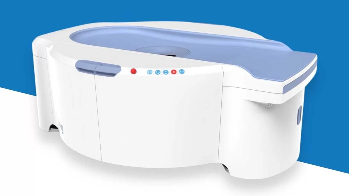
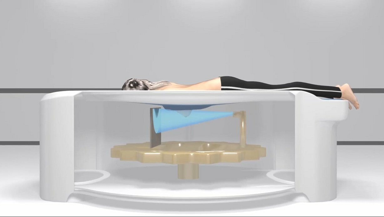
Developed by Georgia-based imaging technology company Koning Health, the Koning Vera Breast CT produces high-resolution, 3D photos in seven seconds and doesn’t compress the breasts the way a mammogram does — making for a more comfortable patient experience and allowing providers to identify tumors with greater accuracy.
The device is particularly helpful in detecting and diagnosing cancer in women with dense breast tissue. Having dense breasts modestly increases one’s likelihood of developing breast cancer, and also makes it harder to spot the disease via traditional mammograms.
This July, the Breast CT was installed at the Bedford Breast Center in Beverly Hills, California, and in April, it made its New York City debut at Community Radiology NY in Manhattan. It’s also currently available for clinical use in Arkansas, Alabama, Georgia, Tennessee, and in parts of Europe and Asia, with installations planned for Texas, Illinois, Florida, and Ohio in the coming months. Use this interactive map to find a location.
“We are thrilled to see our technology being embraced by health care providers globally,” Lutao Ning, CEO of Koning Health, said in a statement. “Our recent and upcoming installations are a clear indication of the impact the Koning Vera is making in the fight against breast cancer. We remain dedicated to pushing the boundaries of what’s possible in breast imaging and ensuring that more women have access to the lifesaving diagnostics they deserve.”
Compared to digital breast tomosynthesis, or DBT, which is a technologically advanced type of mammography and considered by some to be emerging as the gold standard in screening, the Breast CT provides even clearer, more detailed imagery. While DBT is also referred to as a “3D mammogram,” it actually builds those images by layering 2D photos.
“For an image to be true 3D it must be ‘isotropic’ (the same from any angle),” Koning Vera explains on its website. “Breast CT is true 3D — the image can be viewed from any angle equally, which eliminates any overlapping structures.”
The first prototype of the device — which has a radiation dose in the same range as diagnostic mammography — was created in 2006 and clinical trials commenced the same year. In 2017, the Food and Drug Administration approved it for commercial use, and three years later, the American Medical Association recognized the device with six CPT codes, enabling providers to bill health insurance companies for its use.
Note: Mammograms remain an essential tool in detecting breast cancer. Read up on the American Cancer Society’s recommendations and learn how to perform a breast self-exam.
RELATED: All Women Over 40 Should Be Getting Mammograms, New Breast Cancer Guidelines Urge — What to Know