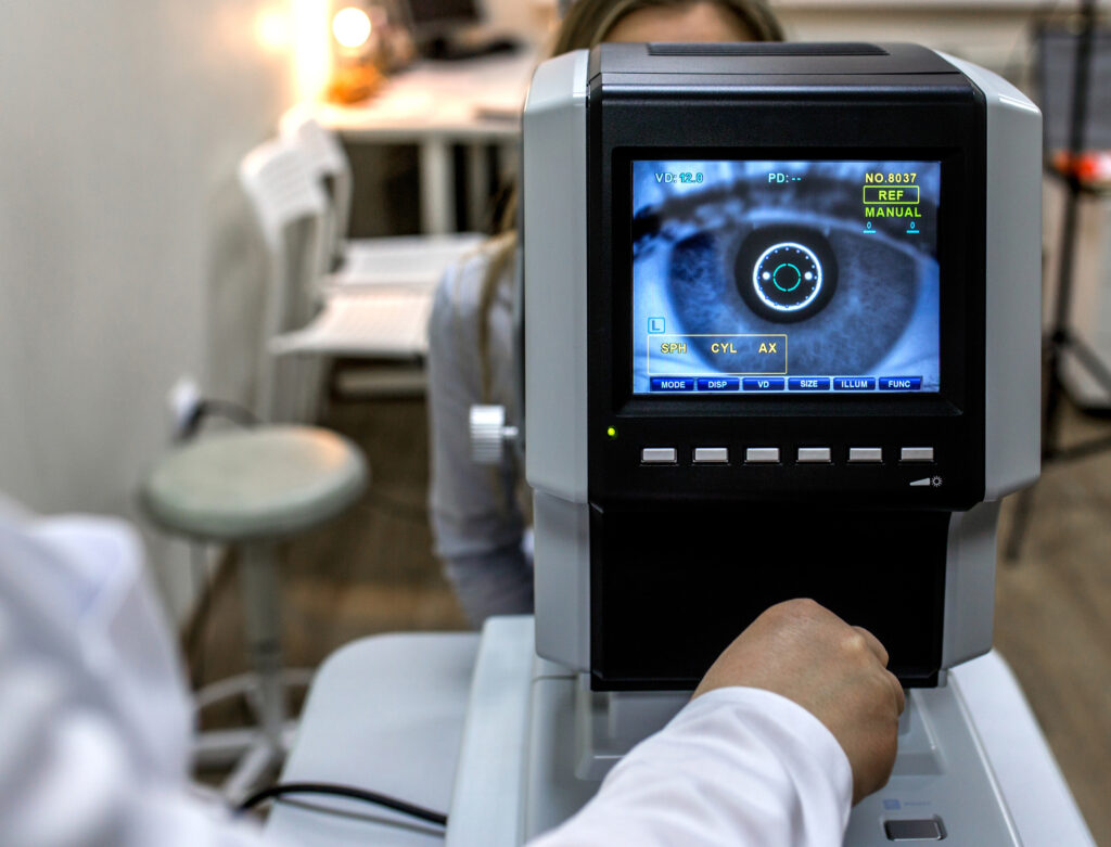
For over a decade, a team of scientists from the University of Wisconsin-Madison have been developing lab-grown retinal neurons with the goal of restoring vision to people with certain degenerative eye diseases. Now, breakthrough testing results suggest that those cells are finally ready to be introduced to patients’ eyes for the first time in clinical trials.
Called retinal organoids (ROs), the neurons are derived from human skin cells that have been programmed to act as stem cells, which can self-replicate. In previous research, cone photoreceptors from these ROs were shown to both detect light and convert the signal into an electric wave — a key element in vision function. According to the National Institutes of Health, in degenerative eye disorders, healthy cells responsible for capturing light and sending visual information can die off, and the body is then unable to regenerate them.

“For diseases like macular degeneration, where cones in the central-most part of the retina die, causing blindness, there are currently no treatment options,” UW–Madison neuroscience professor Raunak Sinha said in a statement to the institution last February. “But with the advent of stem cell technology, you can make these stem cells grow into three-dimensional mini retinas containing cones that can replicate the physiology and function of foveal cones.”
For the lab-grown neurons to work, though, their cone photoreceptors must be able to connect with adjacent cells by forming synapses. Researchers published evidence in May that the cones are able to extend the biological cords, called axons, necessary to make those connections, but until now, there had been no direct proof that they could complete the process.
David Gamm, whose lab developed the organoids, is the director of UW-Madison’s McPherson Eye Research Institute and the co-author of a new study published on January 4. “The last piece of the puzzle was to see if these cords had the ability to plug into, or shake hands with, other retinal cell types in order to communicate,” he said in a news release from the university.
And “shake hands” they did. Using a method called “synaptic tracing,” Gamm and his team were able to verify that cells that had been exposed to a virus had infected adjacent cells with the same virus via synaptic connections.
“We’ve been quilting this story together in the lab, one piece at a time, to build confidence that we’re headed in the right direction,” said Gamm, who co-founded Madison-based Opsis Therapeutics to adapt the technology for the treatment of eye disorders. “It’s all leading, ultimately, to human clinical trials, which are the clear next step.”
He added: “That was an important revelation for us. It really shows the potentially broad impact these retinal organoids could have.”


Pingback: Be My Eyes: Popular App Tests “Life-Altering” AI Tool to Help Visually Impaired People - Usernames Ideas
Pingback: https://peakybet.com/blog
Pingback: เว็บหวยพันธุ์แท้ จ่ายพัน jay1000
Pingback: iux รีวิว
Pingback: chicken road 2 deutschland
Pingback: ร้านต่อผม
Pingback: Stem Cell