Science’s coolest moments aren’t always visible to the naked eye, but the University of Wisconsin-Madison’s appropriately named Cool Science Image Contest is here to show them to us. Featuring objects and phenomena captured through microscopy, photography, animations, medical imaging, and other methods, the competition highlights the best scientific visuals to come out of research, scholarship, or self-guided discovery in the university’s community.
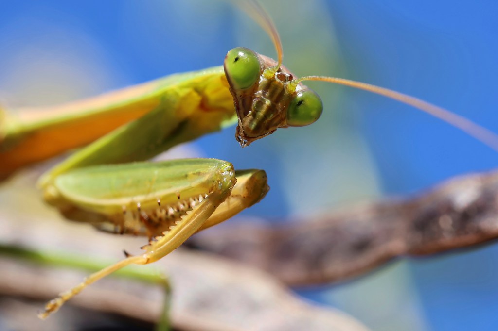
This year’s 13 winners, announced Oct. 1, include the above close-up of a Chinese mantis seemingly giving some serious side-eye. Captured with a digital camera by Kai Skadahl, a financial specialist at the school’s Space Science and Engineering Center, the image actually depicts an optical illusion in the black dots that appear to be the critter’s pupils.
“Each mantis eye is covered in thousands of cells with their own bundle of photoreceptors — like your retina, but covering the outside of the eye,” the caption explains. “The groups of cells you are looking at head-on are not reflecting light back at you, making a dark spot that looks like the pupil in your eye.”
Other standouts of the contest, now in its 15th year, include an embryo of an Arizona bark scorpion that bears an odd resemblance to Star Wars’ Jabba the Hutt, a microscopic shot of a Northern white cedar tree’s leaves, and a photo featuring Lake Michigan’s clear waters. The winners were recognized at an Oct. 3 reception that kicked off an exhibit featuring their images, which runs through January at the McPherson Eye Research Institute’s Mandelbaum and Albert Family Vision Gallery in Madison’s Wisconsin Institutes for Medical Research.
Scroll down to see the other winning shots.
Prashant P. Sharma, Professor of Integrative Biology
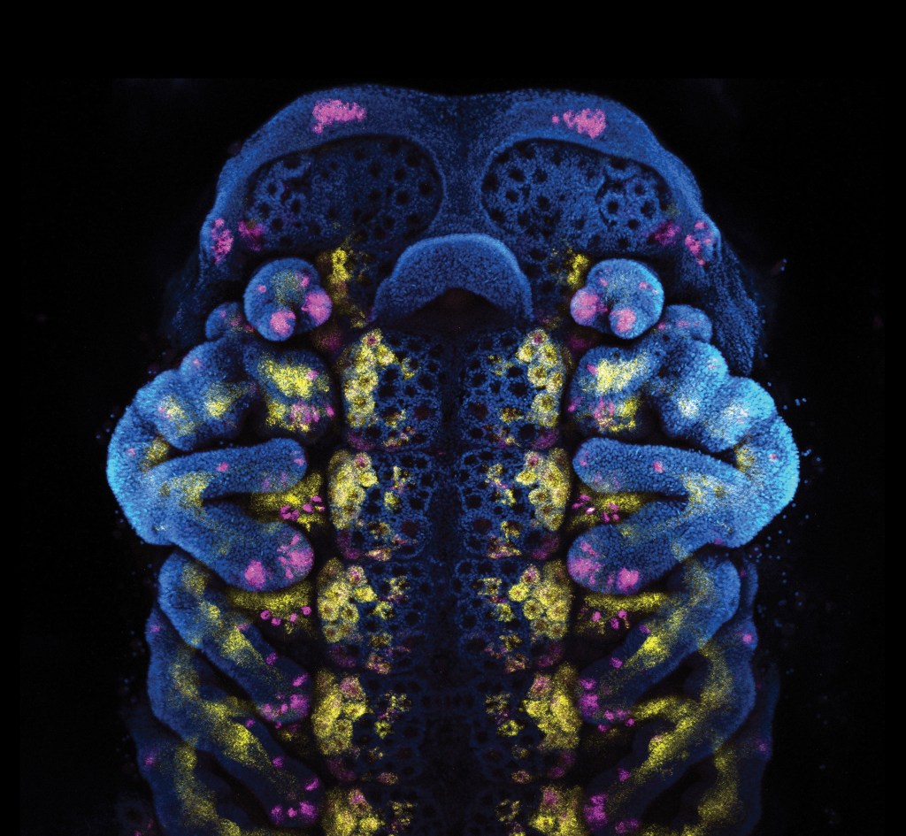
Confocal microscope
“This teddy bear won’t be cuddly for long. It’s the embryo of an Arizona bark scorpion. Spots of color map the expression of two different copies a gene that play an important role in nervous system development. The separate expression patterns show researchers how the respective copies’ functions have diverged over millions of years of evolution.”
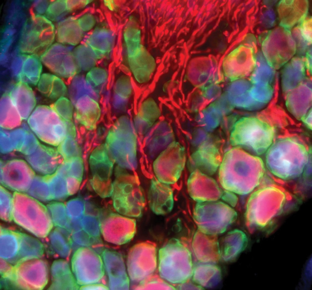
Emily Tran, Graduate Student, Comparative Biomedical Sciences
Tissue imaging fluorescent microscope
“Each unique color marks a thready cell in tissue from a mouse bladder as a different type of sensory neuron, reporting different conditions — stretching, contraction, pain — to the animal’s nervous system. By sorting out the types of neurons at play, researchers hope to improve treatment of bladder pain in humans.”
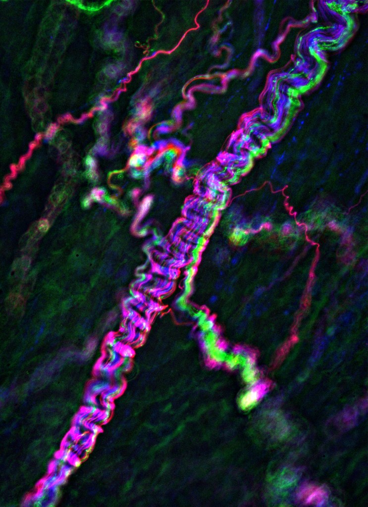
Emily Tran, Graduate Student, Comparative Biomedical Sciences
LaTasha Crawford, Assistant Professor, Pathobiological Sciences
Tissue imaging fluorescent microscope
“A bundle of mouse nerve cells (with different types differentiated by color) are wrapped in the green of signal-regulating cells called satellite glia. In mammals, bundled neurons like these carry signals from the red cells, called axons, of the extended nervous system to the brain.”
Duncan D. Smith, Scientist, Botany
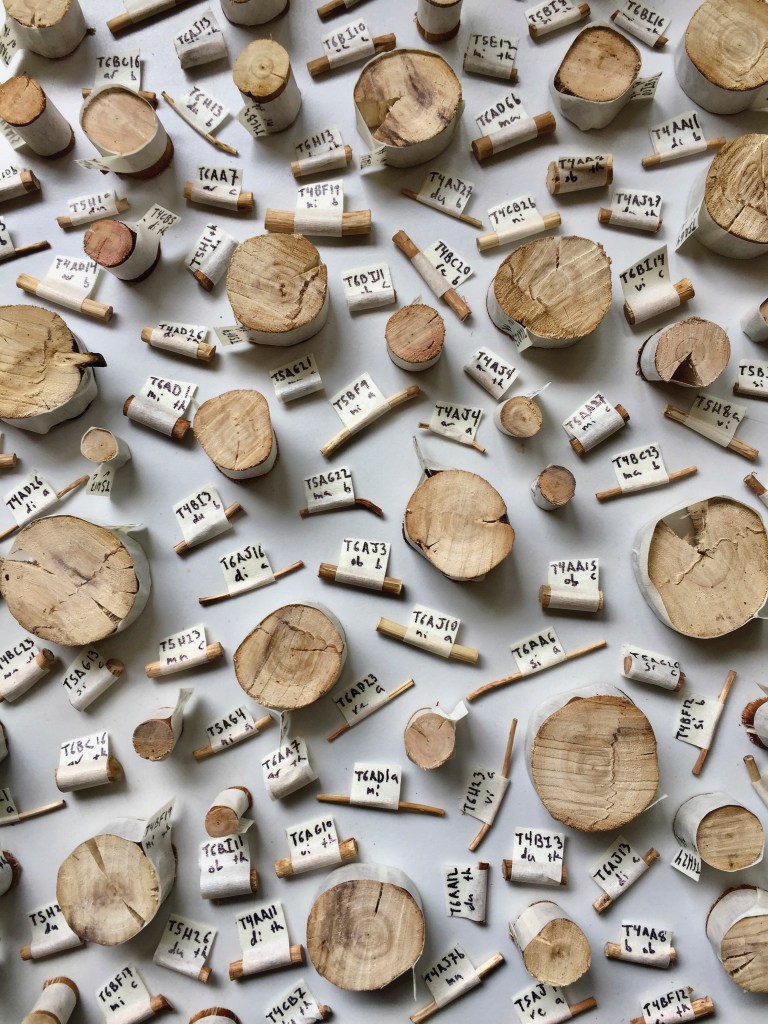
Digital camera
“Measurements of density and descriptions of cellular anatomy derived from these eucalyptus samples collected across contrasting sites in Australia help researchers understand how differences in moisture levels affect the way trees invest in the structure of their wood.”
Christine A. Clark, Outreach Specialist, Division of Extension
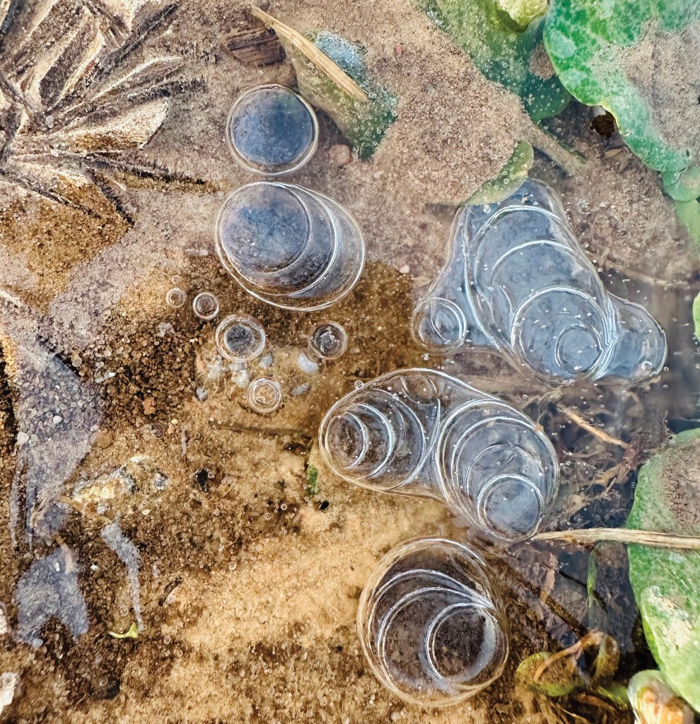
Digital camera
“On an early spring morning in a Wisconsin agricultural field, time appears frozen atop the soil — belying the significant role of spring melt in erosion. Often by midday, this ice melts and flows across ground still frozen beneath the surface, carrying vital topsoil away as it goes.”
Cole Dunbar, Graduate Student, Nuclear Engineering and Engineering Physics
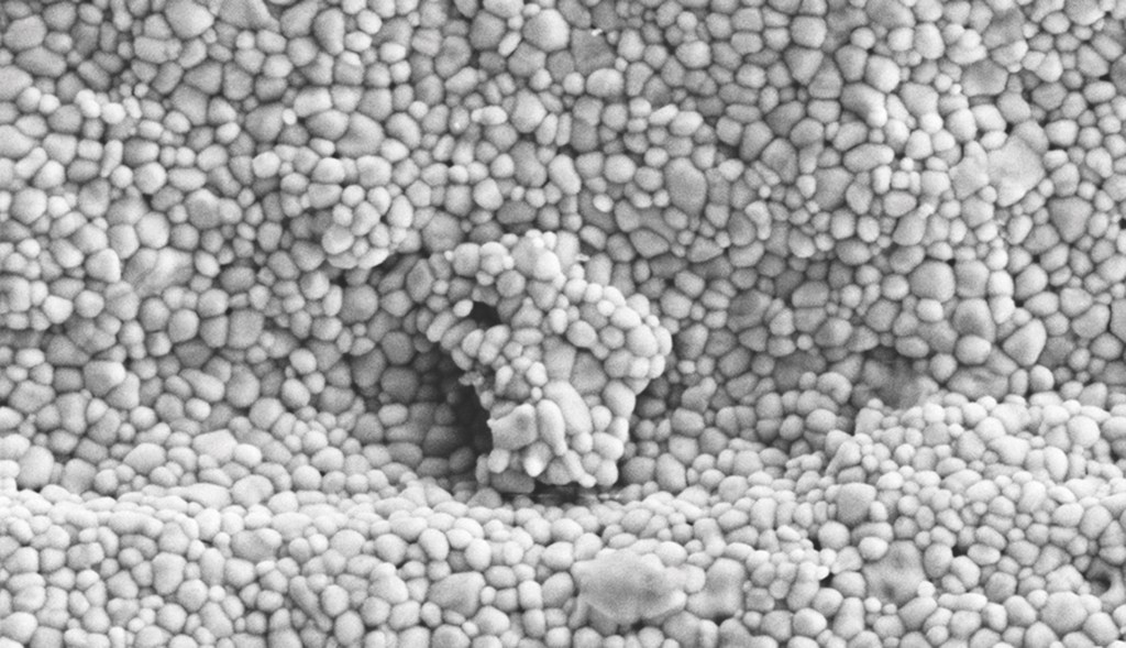
Scanning electron microscope
“Oxidation pebbles the surface of a cladding tube, a layer separating fuel rods from coolant in a nuclear reactor, including a 400-nanometer lump resembling the state of Wisconsin. UW-Madison researchers study wear on cladding to explain changes in qualities like heat transfer during potential accident conditions.”
Shreya Kotha, Research Intern, Mechanical Engineering
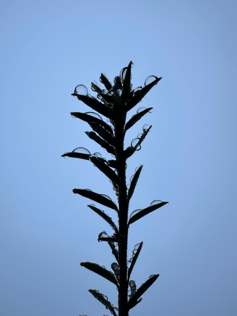
Digital camera
“Droplets of water clinging to a plant after a gentle rain are like tiny physics labs — focusing light, magnifying the textures of the leaves beneath and demonstrating the surface tension holding the water molecules in tidy domes against the pull of gravity.”
Eleanor Ford, Research Intern, Cellular and Molecular Biology

CT scan
“The trachea (or windpipe, traced here in yellow) of the alpine ground beetle is relatively unique in every individual, as it is grown as-needed in reaction to low-oxygen conditions during development.”
Lena Berry, Graduate Student, Botany
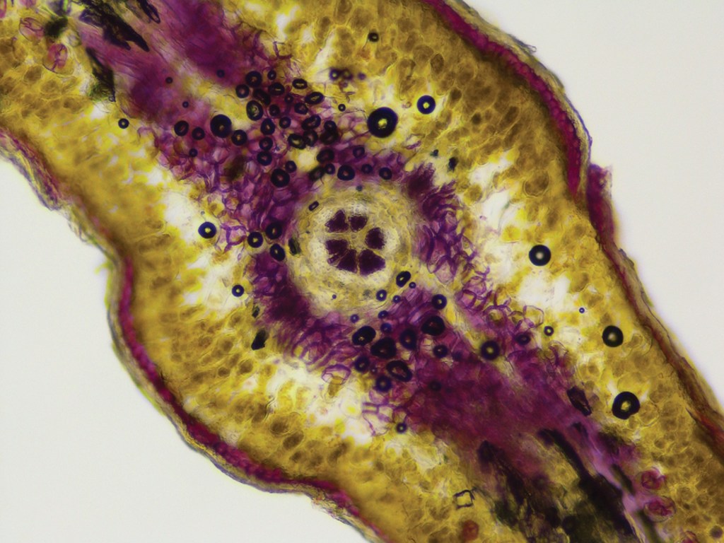
Microscope
“In cross section, the scaly leaves of the Northern white cedar tree are a world of complexity. Cells and cell walls containing stiffening lignin are stained purple, giving researchers the contrast they need to study structures called transfusion tracheids, and how differences in their anatomy make the leaves more or less efficient at moving water.”
Nicholas Hagopian and Yangchen He, Graduate Students, Materials Science and Engineering
Daniel Rhodes and Paul Voyles, Professors, Materials Science and Engineering

Scanning transmission electron microscope
“Each dot in this cross-section of sheets of two-dimensional crystals of tungsten diselenide, WSe2, represents a column of atoms — tungsten in yellow, selenium in pink. Each sheet is less than a nanometer thick. One layer, in the center, was inserted by UW–Madison researchers to disrupt the stack and help them answer questions about the relationships between material properties and atomic structures.”
Matthew Aleksey, Graduate Student, Geoscience
Bil Schneider, Researcher, Geoscience
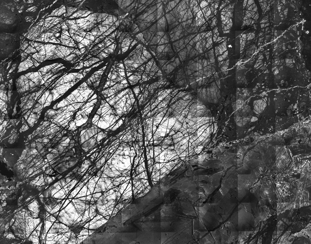
Scanning electron microscope
“This sample of quartz from within an ancient fault in the Baraboo Hills is shot through with microscopic veins of newer quartz formed after tectonic activity cracked the original quartz’s crystal structure. The progressive cracking and overlaying of new veins — like growth rings in a tree — can act as a record the tectonic history of the 1.4-billion-year-old geological fault line.”
Kerry J. Grosse, Policy Analyst, Office of Strategic Consulting

Digital camera
“The clarity of Lake Michigan waters near Coast Guard Station Sturgeon Bay is largely the result of filter-feeding invasive mussels, one way in which the lake’s neighbors have come to understand the way they can affect the life of a body of water that gives back so much — from drinking water and fishing to shipping, recreation and even weather.”


")