This article was originally written by Dean Murray for SWNS — the U.K.’s largest independent news agency, providing globally relevant original, verified, and engaging content to the world’s leading media outlets.
Scientists at the University of Cambridge’s Microscopy Bioscience Platform, a hub uniting advanced imaging facilities across seven departments, has unveiled work that’s helping revolutionize biological research. Through state-of-the-art microscope technologies, the team is unlocking insight into topics ranging from cancer to coral reefs.
Helmed by Lisa-Maria Needham, the platform supports researchers with powerful electron and light microscopes, custom-built tools like “optical tweezers” — which use focused light to manipulate cells — and a dedicated arm for developing new technologies.
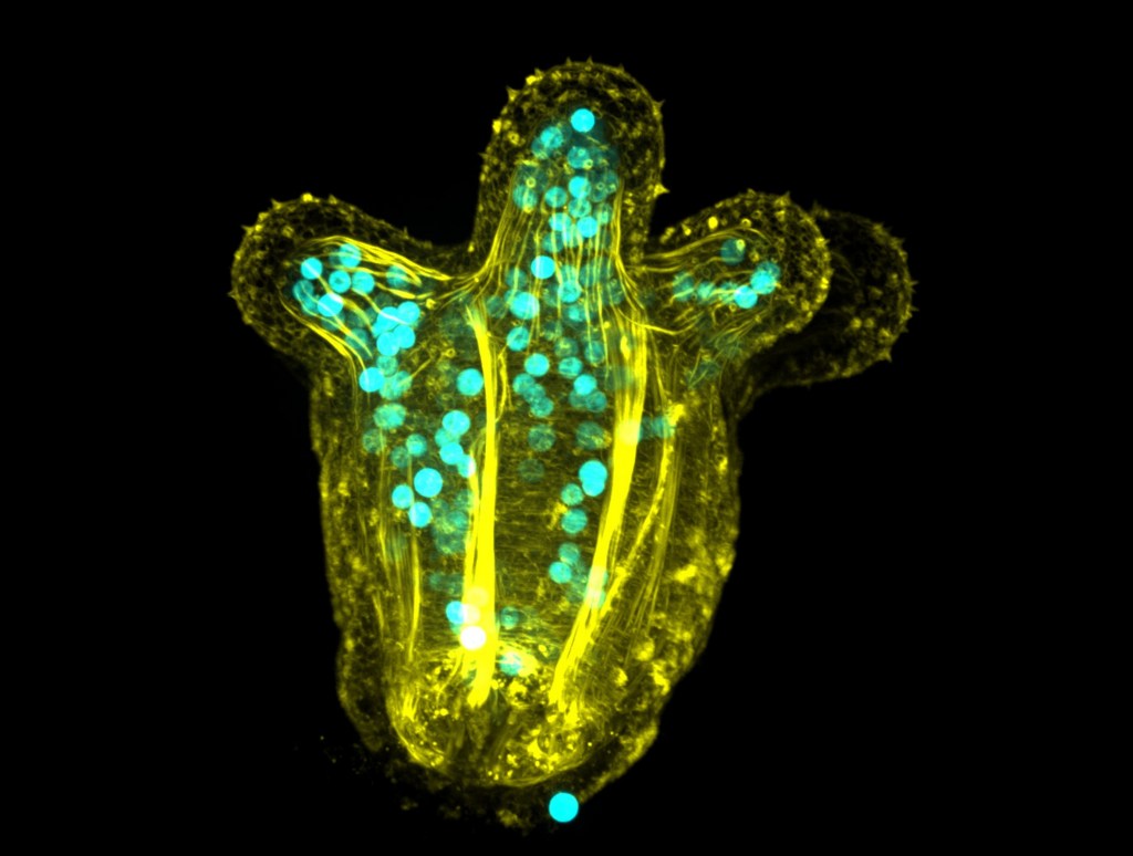
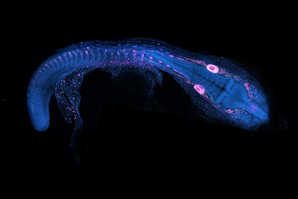
“A developing sea anemone with spherical algae living inside its cells. The tentacles face upwards towards the light and the algae produce nutrients from photosynthesis, transferring some of these nutrients to their host.”
“We’re developing microscopes that don’t exist anywhere in the world,” Needham said in a statement, adding: “The photo-manipulation suite is my favorite microscope, it’s a bit like a Swiss Army knife! It can manipulate biological samples in so many precise ways, helping researchers gain deeper insights into biological processes, diseases, and drug pathways.”
Recent breakthroughs include using live 3D time-lapse microscopy to reveal how animal embryos develop, deploying nanorobots to investigate how cells sense their environment and become cancerous, and studying coral-algae relationships to predict coral bleaching.
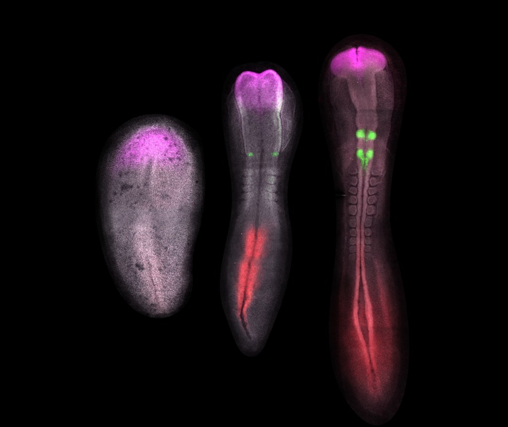
“Chicken embryos at 24 hours, 30 hours, and 38 hours of development, stained with fluorescent dyes to show when different genes are switched on in the developing nervous system. The forebrain (in magenta) develops first, then the hindbrain (in green) and spinal cord (in red).”
Researchers are also uncovering how new fish species emerge through advanced imaging and genetics, and identifying how pancreatic cancer cells survive in harsh conditions to inform new treatments.
“I believe that advances in technology go hand in hand with biological discovery,” said Needham. “Scientists get to a certain point in their research where they know there’s something going on, but nothing exists to help them see it. That’s driving technological innovation.”

“This spinning disk confocal microscopy image reveals cells (blue) growing with nanorobots (red dots).“
Needham wants the platform to become a hub for collaboration, allowing researchers to connect with others across the university and beyond to advance their work. Some of the technologies developed have already been transferred to commercial use through spin-out companies and industrial collaboration.
Take a closer look at the team’s work.


")


")